Canine glaucoma
Glaucoma in dogs refers to a group of diseases that affect the optic nerve and involves a loss of retinal ganglion cells in a characteristic pattern. Raised intraocular pressure is a significant risk factor for developing glaucoma (above 22 mmHg or 2.9 kPa). Untreated glaucoma in dogs leads to permanent damage of the optic nerve and resultant visual field loss, which can progress to blindness.
Canine glaucoma can be divided roughly into two main categories, primary or secondary glaucoma. In dogs, most forms of primary glaucoma are the result of a collapsed filtration angle, or closed angle glaucoma.
Clinical Signs

Dog glaucoma often goes unnoticed until it is in a more severe state. There are rarely any symptoms in the early stages of the disease so regular eye checks by qualified veterinary professionals are important. Dogs will sometimes rub the eye if it is painful. An eye affected with glaucoma may be red, swollen, sore, or become clouded in appearance.
Predisposed Breeds
Certain breeds are predisposed to getting glaucoma. Primary glaucoma most commonly afflicts dogs at 3–7 years of age but can occur at any age. The disease is most frequently seen in cocker spaniels, many of the terrier breeds, Northern breeds such as the Siberian Husky, Poodles, Beagles, chow-chows, jack russell terriers, bassett hounds and Dalmatians. However, primary glaucoma has been identified in almost every breed of dog.
Diagnosis
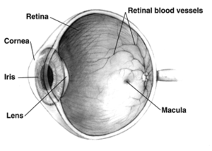
Veterinarians employ three general methods: (1) tonometry, (2) gonioscopy, and (3) ophthalmoscopy. Tonometry measures the IOP with an instrument. Normal IOP in dogs can range between 12 and 25 mm Hg and the two eyes should be similar in pressure. Gonioscopy is a diagnostic procedure to examine the angle of the anterior chamber. Direct and indirect ophthalmoscopy is necessary to evaluate the retina and particularly the optic nerve.
Surgery
Treatment for dog glaucoma has several options, with surgery often being the most effective for long term management.
Laser Surgery
Laser surgery is often performed to selectively destroy the tissue, ciliary body, in an effort to reduce aqueous fluid production. Laser surgery can also be combined with placement of a shunt.
Enucleation
The eyeball is removed during this procedure, often reserved for patients with end stage glaucoma.
Intraocular Evisceration & Implantation
The inner contents of the eye are removed and replaced with an implant. The outer portions of the eye remain.
Canine Specific Intra-Ocular Shunt: TR-ClarifEYE
TR-ClarifEYE is a new implant made of a new biomaterial, the STAR BioMaterial, which consists of silicone with a very precise homogenous pore size, a property which reduces fibrosis and improves tissue integration. The implant contains no valves and is placed completely within the eye without sutures. To date, it has demonstrated long term success (> 1yr) in a pilot study in medically refractory dogs with advanced glaucoma [1]
Valved Shunts
There are also several different glaucoma drainage implants. These include the original Molteno implant (1966), the Baerveldt tube shunt, or the valved implants, such as the Ahmed glaucoma valve implant or the ExPress Mini Shunt and the later generation pressure ridge Molteno implants. These are indicated for glaucoma patients not responding to maximal medical therapy, with previous failed guarded filtering surgery (trabeculectomy). The flow tube is inserted into the anterior chamber of the eye and the plate is implanted underneath the conjunctiva to allow flow of aqueous fluid out of the eye into a chamber called a bleb.
- The first-generation Molteno and other non-valved implants sometimes require the ligation of the tube until the bleb formed is mildly fibrosed and water-tight[2] This is done to reduce postoperative hypotony—sudden drops in postoperative intraocular pressure (IOP).
- Valved implants such as the Ahmed glaucoma valve attempt to control postoperative hypotony by using a mechanical valve.
The ongoing scarring over the conjunctival dissipation segment of the shunt may become too thick for the aqueous humor to filter through. This may require preventive measures using anti-fibrotic medication like 5-fluorouracil (5-FU) or mitomycin-C (during the procedure), or additional surgery. And for Glaucomatous painful Blind Eye and some cases of Glaucoma, Cyclocryotherapy for ciliary body ablation could be considered to be performed.[3]
See also
References
- ↑ Roberts S, Woods C. Effects of a novel porous implant in refractory glaucomatous dogs. ACVO abstract 2008, Boston, MA.
- ↑ Molteno AC, Polkinghorne PJ, Bowbyes JA (November 1986). "The vicryl tie technique for inserting a draining implant in the treatment of secondary glaucoma". Aust N Z J Ophthalmol. 14 (4): 343–54. doi:10.1111/j.1442-9071.1986.tb00470.x. PMID 3814422.
- ↑ Pardianto G; et al. (2006). "Some difficulties on Glaucoma". Mimbar Ilmiah Oftalmologi Indonesia. 3: 49–50.