Calmodulin


Calmodulin (CaM) (an abbreviation for calcium-modulated protein) is a multifunctional intermediate calcium-binding messenger protein expressed in all eukaryotic cells.[1] It is an intracellular target of the secondary messenger Ca2+, and the binding of Ca2+ is required for the activation of Calmodulin. Once bound to Ca2+, Calmodulin acts as part of a calcium signal transduction pathway by modifying its interactions with various target proteins such as kinases or phosphatases.[2][3][4]
Structure
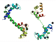
Calmodulin is a small, highly conserved protein that is 148 amino acids long (16.7 KDa). The protein has two approximately symmetrical globular domains each containing a pair of EF-hand motifs (the N- and C-domain) separated by a flexible linker region for a total of four Ca2+ binding sites.[5] Each EF-hand motif allows calmodulin to sense intracellular calcium levels by binding up to four Ca2+ ions. Calcium ion binding regions are found in the following positions in the sequence of amino acids: 21-32, 57-68, 94-105 and 130-141; each region that calcium binds to is exactly 12 amino acids long. These regions are located between two alpha helices in the EF-hand motifs, the first two regions (21-32 and 57-68) are on one side of the linker region the other two (94-105 and 130-141) are on the other side.[4]
Importance of Flexibility in Calmodulin
Calmodulin binds such a wide variety of target proteins, making it especially important for it to have flexibility. Though Calmodulin's flexibility is more evident when it is bound to a target protein, NMR studies have shown that the linker region of Calmodulin is flexible, even when it is not bound to a target protein. Another important characteristic of calmodulin that allows it to bind a large variety of target proteins is the generic shape of the non-polar grooves in the binding sites. Since the non-polar grooves are generic, they don't require the target proteins to have any specific sequence of amino acids allowing a larger variety of target proteins to be bound. Together, these two structural characteristics of calmodulin allow it to flexibly bind target proteins with various shapes and amino acid sequences.[5] For example, calmodulin binds both NMDA receptors and potassium channels which differ in length by about 50 amino acid residues.[6][7]
Similarity to Troponin C
Calmodulin's structure is very similar to the structure of Troponin C (which is another calcium binding protein). They are both members of the EFh super family. Troponin C, like Calmodulin, has two globular domains that are connected by a linker region.[8] However, Troponin C and Calmodulin differ in the length of the linker region; the linker region of Calmodulin is smaller than that of Troponin C.[8] These remarkably similar structures are an example of how the EF hand motif is highly conserved in calcium binding proteins. Though they have similar structures, their functions are very different. Troponin C has a very specific function (to elicit a conformational change in Troponin I) ultimately causing a contraction in skeletal muscles. Calmodulin, evolved to bind a wider variety of target proteins, allowing it to play a role in many physiological events.[5][8]
Mechanism

Overall


Up to four calcium ions are bound by calmodulin via its four EF hand motifs.[5] EF hands supply an electronegative environment for ion coordination. After calcium binding, hydrophobic methyl groups from methionine residues become exposed on the protein via conformational change. Using both X-Ray and NMR studies, scientists were able to determine that the conformational changes occurred in the alpha-helices of the EF motif, which changes the binding affinity for target proteins. When the alpha helices are perpendicular to one another, the Calmodulin is in an open conformation giving it a higher affinity for target proteins.[9] More specifically, this conformational change presents hydrophobic surfaces, which can in turn bind to Basic Amphiphilic Helices (BAA helices) on the target protein. These helices contain complementary hydrophobic regions. The flexibility of calmodulin's hinged region allows the molecule to wrap around its target.[5] This property allows it to tightly bind to a wide range of different target proteins. The C-domain of calmodulin has a higher affinity for calcium than does the N-domain.
Dynamic features
The C-terminal domain solution structure is similar to the X-ray crystal structure, while the EF hands of the N-terminal domain are considerably less open to the X-ray crystal structure. This indicates that Ca2+ binding causes a larger conformational change in the N-domain than in the C-domain. The backbone flexibility within calmodulin is key to its ability to bind a wide range of targets.[10] Protein domains, connected by intrinsically disordered flexible linker domains, induce long-range allostery, or the conformational change of a protein by ligand binding to an allosteric site (a site other than the functional site), via protein domain dynamics.[10]
Function
General role in the body
Calmodulin mediates many crucial processes such as inflammation, metabolism, apoptosis, smooth muscle contraction, intracellular movement, short-term and long-term memory, and the immune response.[11][12] Calcium participates in an intracellular signaling system by acting as a diffusible second messenger to the initial stimuli. It does this by binding various targets in the cell including a large number of enzymes, ion channels, aquaporins and other proteins.[4] Calmodulin is expressed in many cell types and can have different subcellular locations, including the cytoplasm, within organelles, or associated with the plasma or organelle membranes, but it is always found intracellularly.[12] Many of the proteins that Calmodulin binds are unable to bind calcium themselves, and use calmodulin as a calcium sensor and signal transducer. Calmodulin can also make use of the calcium stores in the endoplasmic reticulum, and the sarcoplasmic reticulum. Calmodulin can undergo post-translational modifications, such as phosphorylation, acetylation, methylation and proteolytic cleavage, each of which has potential to modulate its actions.
Specific Examples
Role in smooth muscle contraction

Calmodulin plays an important role in excitation contraction (EC) coupling and the initiation of the cross-bridge cycling in smooth muscle, ultimately causing smooth muscle contraction.[13] In order to activate contraction of smooth muscle, the head of the myosin light chain must be phosphorylated. This phosphorylation is done by Myosin Light Chain (MLC) Kinase. This MLC Kinase is activated by a calmodulin when it is bound by calcium, thus making smooth muscle contraction dependent on the presence of calcium, through the binding of calmodulin and activation of MLC kinase.[13]
Another way that calmodulin affects muscle contraction is by controlling the movement of Ca2+ across both the cell and sarcoplasmic reticulum membranes. The Ca2+ channels, such as the ryanodine receptor of the sarcoplasmic reticulum, can be activated by calmodulin bound to calcium, thus affecting the overall levels of calcium in the cell.[14]
This is a very important function of calmodulin because it indirectly plays a role in every physiological process that is affected by smooth muscle contraction such as digestion and contraction of arteries (which helps distribute blood and regulate blood pressure).[15]
Role in metabolism
Calmodulin plays an important role in the activation of phosphorylase kinase, which cleaves glucose from glycogen.[16]
Calmodulin also plays an important role in lipid metabolism by affecting Calcitonin. Calcitonin is a polypeptide hormone that lowers blood Ca2+ levels and activates G Protein cascades that leads to the generation of cAMP. The actions of calcitonin can be blocked by inhibiting the actions of calmodulin, suggesting that calmodulin plays a crucial role in the activation of calcitonin.[16]
Role in short-term and long-term memory
Long-term potentiation (LTP) requires a depolarization of GABAergic neurons (Neurons that use GABA as a neurotransmitter) in the hippocampus.[17] Calmodulin Kinase II (CaM-K II) plays a crucial role in achieving LTP. It contributes to the phosphorylation of an AMPA receptor which increases the sensitivity of AMPA receptors.[18] Furthermore, research shows that inhibiting CaM-K II interferes with LTP.[18]
Family members
- Calmodulin 1 (CALM1)
- Calmodulin 2 (CALM2)
- Calmodulin 3 (CALM3)
- Calmodulin-like 1 (CALML1)
- Calmodulin-like 3 (CALML3)
- Calmodulin-like 4 (CALML4)
- Calmodulin-like 5 (CALML5)
- Calmodulin-like 6 (CALML6)
Other calcium-binding proteins
Calmodulin belongs to one of the two main groups of calcium-binding proteins, called EF hand proteins. The other group, called annexins, bind calcium and phospholipid (e.g., lipocortin). Many other proteins bind calcium, although binding calcium may not be considered their principal function in the cell.
See also
- Proteopedia page for Calmodulin and its conformational change
- Protein kinase
- Ca2+/calmodulin-dependent protein kinase
References
- ↑ Stevens FC (1983). "Calmodulin: an introduction". Can. J. Biochem. Cell Biol. 61 (8): 906–10. doi:10.1139/o83-115. PMID 6313166.
- ↑ Chin D, Means AR (2000). "Calmodulin: a prototypical calcium sensor". Trends Cell Biol. 10 (8): 322–8. doi:10.1016/S0962-8924(00)01800-6. PMID 10884684.
- ↑ Purves, Dale; Augustine, George; Fitzpatrick, David; Hall, William; LaMantia, Anthony-Samuel; White, Leonard (2012). Neuroscience. Massachusetts: Sinauer Associates, Inc. pp. 95, 147, 148. ISBN 9780878936953.
- 1 2 3 "CALM1 - Calmodulin - Homo sapiens (Human) - CALM1 gene & protein". www.uniprot.org. Retrieved 2016-02-23.
- 1 2 3 4 5 "Calmodulin". pdb101.rcsb.org. Retrieved 2016-02-23.
- ↑ M.A., RCSB Protein Data Bank, Ataman, Z.A., Gakhar, L., Sorensen, B.R., Hell, J.W., Shea,. "RCSB PDB - 2HQW: Crystal Structure of Ca2+/Calmodulin bound to NMDA Receptor NR1C1 peptide Structure Summary Page". www.rcsb.org. Retrieved 2016-03-14.
- ↑ J.F., RCSB Protein Data Bank, Zhang, M., Meng, X.Y., Cui, M., Pascal, J.M., Logothetis, D.E., Zhang,. "RCSB PDB - 4QNH: Calcium-calmodulin (T79D) complexed with the calmodulin binding domain from a small conductance potassium channel SK2-a Structure Summary Page". www.rcsb.org. Retrieved 2016-03-14.
- 1 2 3 "Calmodulin". collab.itc.virginia.edu. Retrieved 2016-02-23.
- ↑ "Calmodulin Target Database". calcium.uhnres.utoronto.ca. Retrieved 2016-02-01.
- 1 2 Chou JJ, Li S, Klee CB, Bax A (November 2001). "Solution structure of Ca(2+)-calmodulin reveals flexible hand-like properties of its domains". Nat. Struct. Biol. 8 (11): 990–7. doi:10.1038/nsb1101-990. PMID 11685248.
- ↑ "Home Page for Calmodulin". structbio.vanderbilt.edu. Retrieved 2016-02-23.
- 1 2 McDowall, Jennifer. "Calmodulin". InterPro Protein Archive. Retrieved 23 February 2016.
- 1 2 Tansey, M. G.; Luby-Phelps, K.; Kamm, K. E.; Stull, J. T. (1994-04-01). "Ca(2+)-dependent phosphorylation of myosin light chain kinase decreases the Ca2+ sensitivity of light chain phosphorylation within smooth muscle cells.". Journal of Biological Chemistry. 269 (13): 9912–9920. ISSN 0021-9258. PMID 8144585.
- ↑ Walsh, M. P. (1994-06-15). "Calmodulin and the regulation of smooth muscle contraction". Molecular and Cellular Biochemistry. 135 (1): 21–41. doi:10.1007/bf00925958. ISSN 0300-8177. PMID 7816054.
- ↑ Martinsen, A; Dessy, C; Morel, N (2014-10-31). "Regulation of calcium channels in smooth muscle: New insights into the role of myosin light chain kinase". Channels. 8 (5): 402–413. doi:10.4161/19336950.2014.950537. ISSN 1933-6950. PMC 4594426
 . PMID 25483583.
 - 1 2 Nishizawa, Y; Okui, Y; Inaba, M; Okuno, S; Yukioka, K; Miki, T; Watanabe, Y; Morii, H (1988-10-01). "Calcium/calmodulin-mediated action of calcitonin on lipid metabolism in rats.". Journal of Clinical Investigation. 82 (4): 1165–1172. doi:10.1172/jci113713. ISSN 0021-9738. PMC 442666. PMID 2844851.
- ↑ Caillard, Olivier; Ben-Ari, Yehezkel; Gaiarsa, Jean-Luc (1999-07-01). "Long-term potentiation of GABAergic synaptic transmission in neonatal rat hippocampus". The Journal of Physiology. 518 (Pt 1): 109–119. doi:10.1111/j.1469-7793.1999.0109r.x. ISSN 0022-3751. PMC 2269393. PMID 10373693.
- 1 2 Lledo, P M; Hjelmstad, G O; Mukherji, S; Soderling, T R; Malenka, R C; Nicoll, R A (1995-11-21). "Calcium/calmodulin-dependent kinase II and long-term potentiation enhance synaptic transmission by the same mechanism.". Proceedings of the National Academy of Sciences of the United States of America. 92 (24): 11175–11179. doi:10.1073/pnas.92.24.11175. ISSN 0027-8424. PMC 40594. PMID 7479960.
External links
- RCSB details...
- Calmodulin at the US National Library of Medicine Medical Subject Headings (MeSH)
- InterPro: IPR015754
- Jennifer McDowall (2003-01-01). "Calmodulin". InterPro Protein Archive. Retrieved 2008-03-22.
- Melanie Nelson; Walter Chazin. "Home Page for Calmodulin". EF-Hand Calcium-Binding Proteins Data Library. Vanderbilt University. Retrieved 2008-03-22.
- Mitsuhiko Ikura. "Calmodulin Target Database". Ontario Cancer Institute, University of Toronto. Retrieved 2008-03-22.