Adenocarcinoma in situ of the lung
| Bronchioloalveolar carcinoma | |
|---|---|
|
Detail of a CT thorax showing pseudonodules with ground glass pattern consistent with bronchioloalveolar carcinoma | |
| Classification and external resources | |
| Specialty | oncology |
| ICD-10 | C34 |
| ICD-9-CM | 162 |
| ICD-O | M8250/3 |
| MeSH | D002282 |

In situ pulmonary adenocarcinoma (AIS), previously called "Bronchioloalveolar carcinoma" (BAC), is a term describing certain variants of lung cancer arising in the distal bronchioles or alveoli that initially exhibit a specific non-invasive growth pattern. BAC is a type of non-small-cell lung cancer (NSCLC). AIS is defined as a small (≤3 cm) solitary tumour with pure alveolar epithelial appearance ("lepidic" growth), lacking any invasion of the interstitium. If completely resected, the prognosis of surgically treated AIS is 100%.[1]
Classification
In WHO-2004, BACs are one of four specific histologic subtypes of lung adenocarcinoma, along with acinar adenocarcinoma, papillary adenocarcinoma, and solid adenocarcinoma with mucin production. However, approximately 80% of adenocarcinomas are found to contain two (or more) of these four subtypes. Multiphasic tumors such as these are classified into a fifth "subtype", termed adenocarcinoma with mixed subtypes.[2]
There are other classification systems that have been proposed for lung cancers, including BACs and other forms of adenocarcinoma. The Noguchi classification system for small adenocarcinomas has received considerable attention, particularly in Japan, but has not been nearly as widely applied and recognized as the WHO system.[3]
Like other forms of lung carcinoma, BAC possesses unique clinical and pathological features, prognosis, and responses to different treatments.[4]
Diagnosis
The criteria for diagnosing BACs have changed since 1999.[5][6] Under the new definition, BAC is defined as a tumor that grows in a lepidic (that is, a scaly covering) fashion along pre-existing airway structures, without detectable invasion or destruction of the underlying tissue, blood vessels, or lymphatics.[7] Because invasion must be ruled out, BAC can be diagnosed only after complete sectioning and examination of the entire tumor, not using biopsy or cytology samples. BAC is considered a pre-invasive malignant lesion that, after further mutation and progression, eventually generates an invasive adenocarcinoma. Therefore, it is considered a form of carcinoma in situ (CIS).
Epidemiology
The criteria for diagnosing BAC have changed since 1999.[7] Under the new definition, BAC is not considered to be an invasive tumor by pathologists, but as one form of carcinoma in situ (CIS). Like other forms of CIS, BAC may progress and become overtly invasive, exhibiting malignant, often lethal, behavior. Major surgery, either a lobectomy or a pneumonectomy, is usually needed to control it, and like other forms of non-small cell lung carcinoma, recurrences are frequent. Therefore, oncologists classify it among the other malignant tumors, which are invasive tumors.[2]
Under the new, more restrictive WHO criteria for lung cancer classification, BAC is now diagnosed much less frequently than it was in the past.[2] Recent studies suggest that BAC comprises between 3% and 5% of all lung carcinomas in the U.S.[8][9]
Variants of BAC
BAC occurs in two major histopathological variants, mucinous BAC (m-BAC, 20–25% of cases)[10][11] and non mucinous BAC (nm-BAC, 75–80% of cases).[11][12] Very rarely, BAC can also occur as a "mixed mucinous and non-mucinous" (or "indeterminant") variant.[2]
Incidence
The incidence of bronchiolo-alveolar carcinoma has been reported to vary from 4–24% of all lung cancer patients.[9] An analysis of Surveillance epidemiology and End results registry ( SEER) by Read et al. revealed that although the incidence of BAC has increased over the past two decade it still constitutes less than 4% of NSCLC in every time interval.[9] This difference in the incidence has been attributed to complex histopathology of cancer. While pure BAC is rare, the increase in incidence as seen in various studies can be due to unclear histological classification till WHO came up with its classification in 1999 and then in 2004. Another distinguishing feature about BAC is that it afflicts men and women in equal proportions, some recent studies even suggest slightly higher incidence among women.[8][9]
Histogenesis
Nonmucinous BAC is thought to derive from a transformed cell in the distal airways and terminal respiratory units, and often shows features of club cell or Type II pneumocyte differentiation.[10] Mucinous BAC, in contrast, probably derives from a transformed glandular cell in distal bronchioles.[13]
Type-I cystic adenomatoid malformation (CAM) has recently been identified as a precursor lesion for the development of mucinous BAC, but these cases are rare.[14][15]
Rarely, BAC may develop a rhabdoid morphology due to the development of dense perinuclear inclusions.[16]
Genetics
The genes mutated in AIS differ based on exposure to tobacco smoke. Non-smokers with AIS commonly have mutations in EGFR (a driver) or HER2 (an important oncogene), or a gene fusion with ALK or ROS1 as one of the elements.[17]
Tumorigenesis
A multi-step carcinogenesis hypothesis suggests a progression from pulmonary atypical adenomatous hyperplasia (AAH) through bronchioalveolar carcinoma (BAC) to invasive adenocarcinoma (AC), but to date this has not been formally demonstrated.[18]
Treatment
The treatment of choice in any patient with BAC is complete surgical resection, typically via lobectomy or pneumonectomy, with concurrent ipsilateral lymphadenectomy.[11]
Non-mucinous BACs are highly associated with classical EGFR mutations, and thus are often responsive to targeted chemotherapy with erlotinib and gefitinib. K-ras mutations are rare in nm-BAC.[19]
Mucinous BAC, in contrast, is much more highly associated with K-ras mutations and wild-type EGFR, and are thus usually insensitive to the EGFR tyrosine kinase inhibitors.[20] In fact, there is some evidence that suggests that the administration of EGFR-pathway inhibitors to patients with K-ras mutated BACs may even be harmful.[21]
Recurrence
When BAC recurs after surgery, the recurrences are local in about three-quarters of cases, a rate higher than other forms of NSCLC, which tends to recur distantly.[11]
Prognosis and survival
Taken as a class, long-term survival rates in BAC tend to be higher than those of other forms of NSCLC.[22][23] BAC generally carries a better prognosis than other forms of NSCLC, which can be partially attributed to localized presentation of the disease.[9] Though other factors might play a role. Prognosis of BAC depends upon the histological subtype and extent at presentation but are generally same as other NSCLC.[24]
Recent research has made it clear that nonmucinous and mucinous BACs are very different types of lung cancer.[10][25] Mucinous BAC is much more likely to present with multiple unilateral tumors and/or in a unilateral or bilateral pneumonic form than nonmucinous BAC.[10] The overall prognosis for patients with mucinous BAC is significantly worse than patients with nonmucinous BAC.[8][26]
Although data are scarce, some studies suggest that survival rates are even lower in the mixed mucinous/non-mucinous variant than in the monophasic forms.[26]
In non-mucinous BAC, neither Clara cell nor Type II pneumocyte differentiation appears to affect survival or prognosis.[10]
Additional images
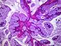
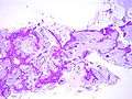
Mucinous BAC
.jpg)
.jpg)
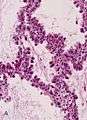
Non-mucinous BAC
See also
- Atypical adenomatous hyperplasia of the lung
- Minimally invasive adenocarcinoma of the lung
- Adenocarcinoma of the lung
References
- ↑ Van Schil, P. E.; Asamura, H; Rusch, V. W.; Mitsudomi, T; Tsuboi, M; Brambilla, E; Travis, W. D. (2012). "Surgical implications of the new IASLC/ATS/ERS adenocarcinoma classification". European Respiratory Journal. 39 (2): 478–86. doi:10.1183/09031936.00027511. PMID 21828029.
- 1 2 3 4 Travis, William D; Brambilla, Elisabeth; Muller-Hermelink, H Konrad; Harris, Curtis C, eds. (2004). Pathology and Genetics of Tumours of the Lung, Pleura, Thymus and Heart (PDF). World Health Organization Classification of Tumours. Lyon: IARC Press. ISBN 92-832-2418-3. Retrieved 27 March 2010.
- ↑ Noguchi M, Morikawa A, Kawasaki M, et al. (June 1995). "Small adenocarcinoma of the lung. Histologic characteristics and prognosis". Cancer. 75 (12): 2844–52. doi:10.1002/1097-0142(19950615)75:12<2844::aid-cncr2820751209>3.0.co;2-#. PMID 7773933.
- ↑ Varlotto JM, Flickinger JC, Recht A, Nikolov MC, DeCamp MM (April 2008). "A comparison of survival and disease-specific survival in surgically resected, lymph node-positive bronchioloalveolar carcinoma versus nonsmall cell lung cancer: implications for adjuvant therapy". Cancer. 112 (7): 1547–54. doi:10.1002/cncr.23289. PMID 18260087.
- ↑ Kreyberg, L.; Liebow, A.A.; Uehlinger, E.A.; World Health Organization. (1967). Histological Typing of Lung Tumours. International histological classification of tumours, no. 1 (1st ed.). Geneva, Switzerland: World Health Organization. OCLC 461784.
- ↑ World Health Organization (1981). Histological Typing of Lung Tumours. International histological classification of tumours, no. 1 (2nd ed.). Geneva, Switzerland: World Health Organization. ISBN 92-4-176101-6. OCLC 476258805.
- 1 2 Travis, W.D.; Colby, T.V.; Corrin, B.; et al.Histological Typing of Lung and Pleural Tumors. International histological classification of tumours, no. 1 (3rd ed.). Berlin: Springer. 1999. ISBN 3-540-65219-1.
- 1 2 3 Zell JA, Ou SH, Ziogas A, Anton-Culver H (November 2005). "Epidemiology of bronchioloalveolar carcinoma: improvement in survival after release of the 1999 WHO classification of lung tumors". J. Clin. Oncol. 23 (33): 8396–405. doi:10.1200/JCO.2005.03.0312. PMID 16293870.
- 1 2 3 4 5 Read WL, Page NC, Tierney RM, Piccirillo JF, Govindan R (August 2004). "The epidemiology of bronchioloalveolar carcinoma over the past two decades: analysis of the SEER database". Lung Cancer. 45 (2): 137–42. doi:10.1016/j.lungcan.2004.01.019. PMID 15246183.
- 1 2 3 4 5 Yousem SA, Beasley MB (July 2007). "Bronchioloalveolar carcinoma: a review of current concepts and evolving issues". Arch. Pathol. Lab. Med. 131 (7): 1027–32. doi:10.1043/1543-2165(2007)131[1027:BCAROC]2.0.CO;2. PMID 17616987.
- 1 2 3 4 Raz DJ, He B, Rosell R, Jablons DM (June 2006). "Current concepts in bronchioloalveolar carcinoma biology". Clin. Cancer Res. 12 (12): 3698–704. doi:10.1158/1078-0432.CCR-06-0457. PMID 16778095.
- ↑ Lee KS, Kim Y, Han J, Ko EJ, Park CK, Primack SL (1 November 1997). "Bronchioloalveolar carcinoma: clinical, histopathologic, and radiologic findings". Radiographics. 17 (6): 1345–57. doi:10.1148/radiographics.17.6.9397450. PMID 9397450.
- ↑ Chilosi M, Murer B (January 2010). "Mixed adenocarcinomas of the lung: place in new proposals in classification, mandatory for target therapy". Arch. Pathol. Lab. Med. 134 (1): 55–65. doi:10.1043/1543-2165-134.1.55. PMID 20073606.
- ↑ Scialpi M, Cappabianca S, Rotondo A, et al. (June 2010). "Pulmonary congenital cystic disease in adults. Spiral computed tomography findings with pathologic correlation and management". Radiol Med. 115 (4): 539–50. doi:10.1007/s11547-010-0467-6. PMID 20058095.
- ↑ Abecasis F, Gomes Ferreira M, Oliveira A, Vaz Velho H (2008). "[Bronchioloalveolar carcinoma associated with congenital pulmonary airway malformation in an asymptomatic adolescent]". Rev Port Pneumol (in Portuguese). 14 (2): 285–90. PMID 18363023.
- ↑ Song DE, Jang SJ, Black J, Ro JY (September 2007). "Mucinous bronchioloalveolar carcinoma of lung with a rhabdoid component—report of a case and review of the literature". Histopathology. 51 (3): 427–30. doi:10.1111/j.1365-2559.2007.02784.x. PMID 17727492.
- ↑ World Cancer Report 2014. World Health Organization. 2014. pp. Chapter 5.1. ISBN 9283204298.
- ↑ Bettio, D; Cariboni, U; Venci, A; Valente, M; Spaggiari, P; Alloisio, M (2012). "Cytogenetic findings in lung cancer that illuminate its biological history from adenomatous hyperplasia to bronchioalveolar carcinoma to adenocarcinoma: A case report". Experimental and Therapeutic Medicine. 4 (6): 1032–1034. doi:10.3892/etm.2012.725. PMC 3494121
 . PMID 23226769.
 - ↑ Finberg KE, Sequist LV, Joshi VA, et al. (July 2007). "Mucinous differentiation correlates with absence of EGFR mutation and presence of KRAS mutation in lung adenocarcinomas with bronchioloalveolar features". J Mol Diagn. 9 (3): 320–6. doi:10.2353/jmoldx.2007.060182. PMC 1899415. PMID 17591931.
- ↑ Sakuma Y, Matsukuma S, Yoshihara M, et al. (July 2007). "Distinctive evaluation of nonmucinous and mucinous subtypes of bronchioloalveolar carcinomas in EGFR and K-ras gene-mutation analyses for Japanese lung adenocarcinomas: confirmation of the correlations with histologic subtypes and gene mutations". Am. J. Clin. Pathol. 128 (1): 100–8. doi:10.1309/WVXFGAFLAUX48DU6. PMID 17580276.
- ↑ Vincent MD (August 2009). "Optimizing the management of advanced non-small-cell lung cancer: a personal view". Curr Oncol. 16 (4): 9–21. doi:10.3747/co.v16i4.465. PMC 2722061. PMID 19672420.
- ↑ Breathnach OS, Kwiatkowski DJ, Finkelstein DM, et al. (January 2001). "Bronchioloalveolar carcinoma of the lung: recurrences and survival in patients with stage I disease". J. Thorac. Cardiovasc. Surg. 121 (1): 42–7. doi:10.1067/mtc.2001.110190. PMID 11135158.
- ↑ Grover FL, Piantadosi S (June 1989). "Recurrence and survival following resection of bronchioloalveolar carcinoma of the lung--The Lung Cancer Study Group experience". Ann. Surg. 209 (6): 779–90. doi:10.1097/00000658-198906000-00016. PMC 1494125. PMID 2543339.
- ↑ Barkley, JE; Green, MR (August 1996). "Bronchioloalveolar carcinoma.". Journal of Clinical Oncology. 14 (8): 2377–86. PMID 8708731.
- ↑ Garfield DH, Cadranel J, West HL (January 2008). "Bronchioloalveolar carcinoma: the case for two diseases". Clin Lung Cancer. 9 (1): 24–9. doi:10.3816/CLC.2008.n.004. PMID 18282354.
- 1 2 Furák J, Troján I, Szoke T, et al. (May 2003). "Bronchioloalveolar lung cancer: occurrence, surgical treatment and survival". Eur J Cardiothorac Surg. 23 (5): 818–23. doi:10.1016/s1010-7940(03)00084-8. PMID 12754039.