Pathogenic bacteria
| Bacterial infection | |
|---|---|
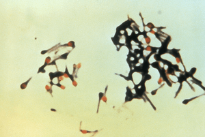 | |
| Clostridium tetani is a pathogenic bacterium that causes tetanus | |
| Classification and external resources | |
| MeSH | D001424 |
Pathogenic bacteria are bacteria that can cause infection. This article deals with human pathogenic bacteria.
Although most bacteria are harmless or often beneficial, several are pathogenic. One of the bacterial diseases with the highest disease burden is tuberculosis, caused by the bacterium Mycobacterium tuberculosis, which kills about 2 million people a year, mostly in sub-Saharan Africa. Pathogenic bacteria contribute to other globally important diseases, such as pneumonia, which can be caused by bacteria such as Streptococcus and Pseudomonas, and foodborne illnesses, which can be caused by bacteria such as Shigella, Campylobacter, and Salmonella. Pathogenic bacteria also cause infections such as tetanus, typhoid fever, diphtheria, syphilis, and leprosy. Pathogenic bacteria are also the cause of high infant mortality rates in developing countries.[1]
Koch's postulates are the standard to establish a causative relationship between a microbe and a disease.
Diseases
Each species has specific effect and causes symptoms in people who are infected. Some, if not most people who are infected with a pathogenic bacteria do not have symptoms. Immuno-compromised individuals are more susceptible to pathogenic bacteria.
Pathogenic susceptibility
Some pathogenic bacteria cause disease under certain conditions, such entry through the skin via a cut, through sexual activity or an compromised immune function.
Streptococcus and Staphylococcus are part of the normal skin microbiota and typically reside on healthy skin or in the nasopharangeal region. Yet these species can potentially initiate skin infections. They are also able to cause sepsis, pneumonia, meningitis. These infections can become quite serious creating a systemic inflammatory response resulting in massive vasodilation, shock, and death.[2]
Other bacteria are opportunistic pathogens and cause disease mainly in people suffering from immunosuppression or cystic fibrosis Examples of these opportunistic pathogens include Pseudomonas aeruginosa, Burkholderia cenocepacia, and Mycobacterium avium.[3][4]
Intracellular
Obligate intracellular parasites (e.g. Chlamydophila, Ehrlichia, Rickettsia) have the ability to only grow and replicate inside other cells. Even these intracellular infections may be asymptomatic, requiring an incubation period. An example of this Rickettsia which causes typhus. Another causes Rocky Mountain spotted fever.
Chlamydia is a phylum of intracellular parasites. These pathogens can cause pneumonia or urinary tract infection and may be involved in coronary heart disease.[5]
Other groups of intracellular bacterial pathogens include: Salmonella, Neisseria, Brucella, Mycobacterium, Nocardia, Listeria, Francisella, Legionella, and Yersinia pestis. These can exist intracellularly, but can exist outside of host cells.
Infections in specific tissue
Bacterial pathogens often cause infection in specific areas of the body. Others are generalists.
- Bacterial vaginosis is caused by bacteria that change the vaginal microbiota caused by an overgrowth of bacteria that crowd out the Lactobacilli species that maintain healthy vaginal microbial populations.
- Other non-bacterial vaginal infections include: yeast infection (candidiasis), Trichomonas vaginalis (trichomoniasis).[6][7]
- Bacterial meningitis is a bacterial inflammation of the meninges, that is, the protective membranes covering the brain and spinal cord.
- Bacterial pneumonia is a bacterial infection of the lungs.
- Urinary tract infection is predominantly caused by bacteria. Symptoms include the strong and frequent sensation or urge to urinate, pain during urination, and urine that is cloudy.[8] The main causal agent is Escherichia coli. Urine is typically sterile but contains a variety of salts, and waste products.[9] Bacteria can ascend into the bladder or kidney and causing cystitis and nephritis.
- Bacterial gastroenteritis is caused by enteric, pathogenic bacteria. These pathogenic species are usually distinct from the usually harmless bacteria of the normal gut flora. But a different strain of the same species may be pathogenic. The distinction is sometimes difficult as in the case of Escherichia.
- Bacterial skin infections include:
- Impetigo is a highly contagious bacterial skin infection commonly seen in children.[10] It is caused by Staphylococcus aureus, and Streptococcus pyogenes.[11]
- Erysipelas is an acute streptococcus bacterial infection[12] of the deeper skin layers that spreads via with lymphatic system.
- Cellulitis is a diffuse inflammation[13] of connective tissue with severe inflammation of dermal and subcutaneous layers of the skin. Cellulitis can be caused by normal skin flora or by contagious contact, and usually occurs through open skin, cuts, blisters, cracks in the skin, insect bites, animal bites, burns, surgical wounds, intravenous drug injection, or sites of intravenous catheter insertion. In most cases it is the skin on the face or lower legs that is affected, though cellulitis can occur in other tissues.
Mechanisms
Nutrients
Iron is required for humans, as well as the growth of most bacteria. To obtain free iron, some pathogens secrete proteins called siderophores, which take the iron away from iron-transport proteins by binding to the iron even more tightly. Once the iron-siderophore complex is formed, it is taken up by siderophore receptors on the bacterial surface and then that iron is brought into the bacterium.[14]
Direct damage
Once pathogens attach to host cells, they can cause direct damage as the pathogens use the host cell for nutrients and produce waste products. As pathogens multiply and divide inside host cells, the cells usually rupture and the intercellular bacteria are released. Some bacteria such as E. coli, Shigella, Salmonella, and Neisseria gonorrhoeae, can induce host epithelial cells to engulf them in a process resembling phagocytosis. The pathogens can then disrupt host cells as they pass through them and be extruded from host cells by a reverse phagocytosis process, enabling them to enter other host cells. Some bacteria can also penetrate host cells by excreting enzymes and by their own motility; such penetration can itself damage the host cell.[14]
Toxin production
Toxins are poisonous substances that are produced by certain microorganisms and are often the primary factor contributing to the pathogenic properties of the microorganisms. Endotoxins are the lipid portions of lipopolysaccharides that are part of the outer membrane of the cell wall of gram negative bacteria. Endotoxins are released when the bacteria lyses, which is why after antibiotic treatment symptoms can at first worsen as the bacteria are killed and they release their endotoxins. Exotoxins are proteins produced inside pathogenic bacteria as part of their growth and metabolism, most common in gram positive bacteria. The exotoxins are released when the bacteria die and the cell wall breaks apart. Exotoxins are highly specific in the effects on body tissues and work by destroying particular parts of the host cell or by inhibiting certain metabolic functions. Exotoxins are among the most lethal known substances, only 1 mg of the botulinum exotoxin is enough to kill one million guinea pigs. Diseases caused this way are often caused by minute amounts of exotoxins, not by the bacteria themselves.[14]
Identification
Typically identification is done by growing the organism in a wide range of cultures which can take up to 48 hours. The growth is then visually or genomically identified. The cultured organism is then subjected to antibiotics to observe reaction to help further identify species and strain.[15]
Treatment
- See also overview list below
Bacterial infections may be treated with antibiotics, which are classified as bacteriocidal if they kill bacteria or bacteriostatic if they just prevent bacterial growth. There are many types of antibiotics and each class inhibits a process that is different in the pathogen from that found in the host. For example, the antibiotics chloramphenicol and tetracyclin inhibit the bacterial ribosome but not the structurally different eukaryotic ribosome, so they exhibit selective toxicity.[16] Antibiotics are used both in treating human disease and in intensive farming to promote animal growth. Both uses may be contributing to the rapid development of antibiotic resistance in bacterial populations.[17] Phage therapy can also be used to treat certain bacterial infections.[18] Infections can be prevented by antiseptic measures such as sterilizing the skin prior to piercing it with the needle of a syringe and by proper care of indwelling catheters. Surgical and dental instruments are also sterilized to prevent infection by bacteria. Disinfectants such as bleach are used to kill bacteria or other pathogens on surfaces to prevent contamination and further reduce the risk of infection. Bacteria in food are killed by cooking to temperatures above 73 °C (163 °F).
List of genera of pathogenic bacteria and microscopy features
Many genera contain pathogenic bacteria species. They often possess characteristics that help to classify and organize them into groups. The following is a partial listing.
| Genus | Species | Gram staining | Shape | Oxygen requirement | Intra/Extracellular |
|---|---|---|---|---|---|
| Bacillus[19] | Positive | Rods | Facultative anaerobic | Extracellular | |
| Bartonella[19] | Negative | Rods | Aerobic | Facultative intracellular | |
| Bordetella[19] | Negative | Small coccobacilli | Aerobic | Extracellular | |
| Borrelia[19] | Negative, stains poorly | spirochete | Anaerobic | Extracellular | |
| Brucella[19] | Negative | coccobacilli | Aerobic | Intracellular | |
| Campylobacter[19] | Negative | Spirochete Bacillus |
microaerophilic | extracellular | |
| Chlamydia and Chlamydophila[19] | (not Gram-stained) | Small, round, ovoid | Facultative or strictly aerobic | Obligate intracellular | |
| Clostridium[19] | Positive | Large, blunt-ended rods | Obligate anaerobic | extracellular | |
| Corynebacterium[19] | Positive (unevenly) | bacilli | Mostly facultative anaerobic | extracellular | |
| Enterococcus[21][24] | Positive | Cocci | Facultative Anaerobic | extracellular | |
| Escherichia[1][21][25] | Negative | Bacillus | Facultative anaerobic | extracellular or intracellular | |
| Francisella[19] | Negative | coccobacillus | strictly aerobic | Facultative intracellular | |
| Haemophilus | Negative | coccobacilli to long and slender filaments | extracellular | ||
| Helicobacter | Negative | Spirochete | Microaerophile | extracellular | |
| Legionella[19] | Negative, stains poorly | cocobacilli | aerobic | facultative intracellular | |
| Leptospira[21][28] | Negative, stains poorly | Spirochete | Strictly aerobic | extracellular | |
| Listeria[19] | Positive, darkly | Slender, short rods | Facultative Anaerobic | intracellular | |
| Mycobacterium[19] | (none) | Long, slender rods | aerobic | extracellular | |
| Mycoplasma[19] | (none) | 'fried egg' appearance, no cell wall | Mostly facultative anaerobic; M. pneumoniae strictly aerobic | extracellular | |
| Neisseria[21][29] | Negative | Kidney bean-shaped | aerobic | Gonococcus: facultative intracellular N. meningitidis: extracellular | |
| Pseudomonas[21][30] | Negative | rods | Obligate aerobic | extracellular | |
| Rickettsia[19] | Negative, stains poorly | Small, rod-like coccobacillary | Aerobic | Obligate intracellular | |
| Salmonella[19] | Negative | Bacillus shape | Facultative anaerobica | Facultative intracellular | |
| Shigella[21][31] | Negative | rods | Facultative anaerobic | extracellular | |
| Staphylococcus[1] | Positive, darkly | Round cocci | Facultative anaerobic | extracellular, facultative intracellular | |
| Streptococcus[19] | Positive | ovoid to spherical | Facultative anaerobic | extracellular | |
| Treponema[19] | Negative, stains poorly | Spirochete | Aerobic | extracellular | |
| Ureaplasma[1] | Stains poorly[32] | indistinct, 'fried egg' appearance, no cell wall | anaerobic | extracellular | |
| Vibrio[21][21][33] | Negative | Spiral with single polar flagellum | Facultative anaerobic | extracellular | |
| Yersinia[21][34] | Negative, bipolarly | Small rods | Facultative Anaerobe | Intracellular | |
List of species of pathogenic bacteria and clinical characteristics
This is description of the more common genera and species presented with their clinical characteristics and treatments.
See also
References
- 1 2 3 4 Santosham, Mathuram; Chan, Grace J.; Lee, Anne CC; Baqui, Abdullah H.; Tan, Jingwen; Black, Robert E. (2013). "Risk of Early-Onset Neonatal Infection with Maternal Infection or Colonization: A Global Systematic Review and Meta-Analysis". PLoS Medicine. 10 (8): e1001502. doi:10.1371/journal.pmed.1001502. ISSN 1549-1676. PMC 3747995
 . PMID 23976885.
 - ↑ Fish DN (February 2002). "Optimal antimicrobial therapy for sepsis". Am J Health Syst Pharm. 59 (Suppl 1): S13–9. PMID 11885408.
- ↑ Heise E (1982). "Diseases associated with immunosuppression". Environ Health Perspect. 43: 9–19. doi:10.2307/3429162. JSTOR 3429162. PMC 1568899. PMID 7037390.
- ↑ Saiman L (2004). "Microbiology of early CF lung disease". Paediatr Respir Rev. 5 (Suppl A): S367–9. doi:10.1016/S1526-0542(04)90065-6. PMID 14980298.
- ↑ Belland R, Ouellette S, Gieffers J, Byrne G (2004). "Chlamydia pneumoniae and atherosclerosis". Cell Microbiol. 6 (2): 117–27. doi:10.1046/j.1462-5822.2003.00352.x. PMID 14706098.
- ↑ Terri Warren, RN (2010). "Is It a Yeast Infection?". Retrieved 2011-02-23.
- ↑ Ferris DG, Nyirjesy P, Sobel JD, Soper D, Pavletic A, Litaker MS (March 2002). "Over-the-counter antifungal drug misuse associated with patient-diagnosed vulvovaginal candidiasis". Obstetrics and Gynecology. 99 (3): 419–425. doi:10.1016/S0029-7844(01)01759-8. PMID 11864668.
- ↑ "Urinary Tract Infections". Retrieved 2010-02-04.
- ↑ "Adult Health Advisor 2005.4: Bacteria in Urine, No Symptoms (Asymptomatic Bacteriuria)". Archived from the original on 2007-07-12. Retrieved 2007-08-25.
- ↑ "Impetigo". National Health Service. Page last reviewed: 17/07/2014
- ↑ Kumar, Vinay; Abbas, Abul K.; Fausto, Nelson; & Mitchell, Richard N. (2007). Robbins Basic Pathology (8th ed.). Saunders Elsevier. pp. 843 ISBN 978-1-4160-2973-1
- ↑ "erysipelas" at Dorland's Medical Dictionary
- ↑ "cellulitis" at Dorland's Medical Dictionary
- 1 2 3 Tortota, Gerard (2013). Microbiology an Introduction. ISBN 978-0-321-73360-3.
- ↑ Cassells AC (2012). "Pathogen and biological contamination management in plant tissue culture: phytopathogens, vitro pathogens, and vitro pests". Methods in Molecular Biology. 877: 57–80. doi:10.1007/978-1-61779-818-4_6. PMID 22610620.
- ↑ Yonath A, Bashan A (2004). "Ribosomal crystallography: initiation, peptide bond formation, and amino acid polymerization are hampered by antibiotics". Annu Rev Microbiol. 58: 233–51. doi:10.1146/annurev.micro.58.030603.123822. PMID 15487937.
- ↑ Khachatourians GG (November 1998). "Agricultural use of antibiotics and the evolution and transfer of antibiotic-resistant bacteria". CMAJ. 159 (9): 1129–36. PMC 1229782. PMID 9835883.
- ↑ Keen, E. C. (2012). "Phage Therapy: Concept to Cure". Frontiers in Microbiology. 3. doi:10.3389/fmicb.2012.00238. PMC 3400130. PMID 22833738.
- 1 2 3 4 5 6 7 8 9 10 11 12 13 14 15 16 17 18 Unless else specified in boxes then ref is: Fisher, Bruce; Harvey, Richard P.; Champe, Pamela C. (2007). Lippincott's Illustrated Reviews: Microbiology (Lippincott's Illustrated Reviews Series). Hagerstown, MD: Lippincott Williams & Wilkins. pp. 332–353. ISBN 0-7817-8215-5.
- ↑ Kurzynski TA, Boehm DM, Rott-Petri JA, Schell RF, Allison PE (1988). "Comparison of modified Bordet-Gengou and modified Regan-Lowe media for the isolation of Bordetella pertussis and Bordetella parapertussis". J. Clin. Microbiol. 26 (12): 2661–3. PMC 266968. PMID 2906642.
- 1 2 3 4 5 6 7 8 9 10 11 12 13 14 15 16 17 18 19 20 21 22 23 24 25 26 27 28 29 30 31 32 33 34 35 36 37 38 39 40 41 42 43 44 45 46 47 48 49 50 51 52 53 54 55 56 57 58 59 60 61 62 63 64 65 66 67 68 69 70 71 72 73 74 75 76 77 78 79 80 81 82 83 84 85 86 87 88 89 90 91 92 93 94 95 96 97 98 99 100 101 102 103 104 105 106 107 108 109 110 111 112 113 114 115 116 117 118 119 120 121 122 123 124 125 126 127 128 129 130 131 132 133 134 135 136 137 138 139 140 141 142 143 144 145 146 147 148 149 150 151 152 153 154 155 156 157 158 159 160 161 162 163 164 165 166 167 168 169 170 171 172 173 174 175 176 177 178 179 180 181 182 183 184 185 186 187 188 189 190 191 192 193 194 195 196 197 198 199 200 201 202 203 204 205 206 207 208 209 210 211 212 213 214 215 216 217 218 219 220 221 222 223 224 225 226 227 228 229 230 231 232 233 234 235 236 237 238 239 240 241 242 243 244 245 246 247 248 249 250 251 252 253 254 255 256 257 258 259 260 261 262 263 264 265 266 267 268 269 270 271 272 273 274 275 276 277 278 279 280 281 282 283 284 285 286 287 288 289 290 291 292 293 294 295 296 297 298 299 300 301 302 303 304 305 306 307 308 309 310 311 312 313 Fisher, Bruce; Harvey, Richard P.; Champe, Pamela C. (2007). Lippincott's Illustrated Reviews: Microbiology (Lippincott's Illustrated Reviews Series). Hagerstown, MD: Lippincott Williams & Wilkins. pp. 332–353. ISBN 0-7817-8215-5.
- 1 2 Bowden GHW (1996). Baron S; et al., eds. Actinomycosis in: Baron's Medical Microbiology (4th ed.). Univ of Texas Medical Branch. ISBN 0-9631172-1-1. (via NCBI Bookshelf).
- ↑ Baron, Samuel (1996). Medical Microbiology (4th ed.). University of Texas Medical Branch at Galveston, Galveston, Texas. ISBN 0-9631172-1-1.
- ↑ Rollins, David M. (2000). "BSCI424 Laboratory Media". University of Maryland. Retrieved 2008-11-18.
- ↑ Cain, Donna (January 14, 2015). "MacConkey Agar (CCCCD Microbiology". Collin College.
- ↑ Gunn BA (1984). "Chocolate agar, a differential medium for gram-positive cocci". Journal of Clinical Microbiology. 20 (4): 822–3. PMC 271442. PMID 6490866.
- ↑ Stevenson TH, Castillo A, Lucia LM, Acuff GR (2000). "Growth of Helicobacter pylori in various liquid and plating media". Lett. Appl. Microbiol. 30 (3): 192–6. doi:10.1046/j.1472-765x.2000.00699.x. PMID 10747249.
- ↑ Johnson RC, Harris VG (1967). "Differentiation of Pathogenic and Saprophytic Leptospires I. Growth at Low Temperatures". J. Bacteriol. 94 (1): 27–31. PMC 251866. PMID 6027998.
- ↑ "Thayer Martin Agar (Modified) Procedure" (PDF). University of Nebraska-Medical Center, Clinical Laboratory Science Program. Retrieved 2015-05-03.
- ↑ Allen, Mary E. (2005). "MacConkey Agar Plates Protocols". American Society for Microbiology. Created: 30 September 2005. Last update: 01 April 2013
- ↑ "Hektoen Enteric Agar". Austin Community College District. Retrieved 2015-05-03.
- ↑ Cassell GH, Waites KB, Crouse DT, Rudd PT, Canupp KC, Stagno S, Cutter GR (1988). "Association of Ureaplasma urealyticum infection of the lower respiratory tract with chronic lung disease and death in very-low-birth-weight infants". Lancet. 2 (8605): 240–5. doi:10.1016/s0140-6736(88)92536-6. PMID 2899235.
- ↑ Pfeffer, C.; Oliver, J.D. (2003). "A comparison of thiosulphate-citrate-bile salts-sucrose (TCBS) agar and thiosulphate-chloride-iodide (TCI) agar for the isolation of Vibrio species from estuarine environments". Letters in Applied Microbiology. 36 (3): 150–151. doi:10.1046/j.1472-765X.2003.01280.x. PMID 12581373.
- ↑ "Yersinia pestis" (PDF). Wadsworth Center. 2006.
- 1 2 3 4 5 6 7 8 9 10 11 12 13 14 15 16 17 18 19 20 21 22 23 24 25 26 27 28 29 30 31 32 33 34 35 36 37 38 39 40 41 42 43 44 45 46 47 48 49 50 51 52 53 54 55 56 57 58 59 60 61 62 63 64 65 66 67 68 69 70 71 72 73 74 75 76 77 78 79 80 81 82 83 84 85 86 87 88 89 90 91 92 93 94 95 96 97 98 99 100 101 102 103 104 105 106 107 108 109 110 111 112 113 114 115 116 117 118 119 120 121 122 123 124 125 126 127 128 129 130 131 132 133 134 135 136 137 138 139 140 141 142 143 144 145 146 147 148 149 150 151 152 153 154 155 156 157 158 159 160 161 162 163 164 165 166 167 168 169 170 171 172 173 174 175 176 177 178 179 180 181 182 183 184 185 186 187 188 189 190 191 192 193 194 195 196 197 198 199 200 201 202 203 204 205 206 207 208 209 210 211 212 213 214 215 216 217 218 219 220 221 222 "Bacteria Table" (PDF). Creighton University School of Medicine. Retrieved 2015-05-03.
- ↑ Brook, I (Oct 2008). "Actinomycosis: diagnosis and management.". Southern Medical Journal. 101 (10): 1019–23. doi:10.1097/SMJ.0b013e3181864c1f. PMID 18791528.
- ↑ Mabeza, GF; Macfarlane J (March 2003). "Pulmonary actinomycosis". European Respiratory Journal. ERS Journals Ltd. 21 (3): 545–551. doi:10.1183/09031936.03.00089103. PMID 12662015. Retrieved 2008-07-21.
- ↑ "Anthrax in animals". Food and Agriculture Organization. 2001.
- ↑ "CDC Anthrax Q & A: Treatment". Retrieved 4 April 2011.
- ↑ "FDA approves raxibacumab to treat inhalational anthrax". Retrieved 14 December 2012.
- 1 2 Itzhak Brook (Jan 28, 2014). "Bacteroides Infection Follow-up". Medscape. Retrieved 2015-09-25.
- ↑ "ESCHERICHIA COLI". Public Health Agency of Canada. 2012-04-30. Retrieved 2015-06-02.
- 1 2 "Signs & Symptoms". Centers for Disease Control and Prevention. Page last reviewed: October 26, 2015
- ↑ Ryan, KJ; Ray, CG, eds. (2004). Sherris Medical Microbiology (4th ed.). McGraw Hill. ISBN 0-8385-8529-9.
- ↑ "Klebsiella pneumoniae in Healthcare Settings". Centers for Disease Control and Prevention. Page last reviewed: November 24, 2010. Page last updated: August 27, 2012
- ↑ Institut Pasteur Press Office - Vaccine against shigellosis (bacillary dysentery):a promising clinical trial 15 January 2009. Retrieved on 27 February 2009
- ↑ Levinson, W. (2010). Review of Medical Microbiology and Immunology (11th ed.). pp. 94–9.
- ↑ Zhou D, Han Y, Yang R (2006). "Molecular and physiological insights into plague transmission, virulence and etiology". Microbes Infect. 8 (1): 273–84. doi:10.1016/j.micinf.2005.06.006. PMID 16182593.
- ↑ Wagle PM. (1948). "Recent advances in the treatment of bubonic plague". Indian J Med Sci. 2: 489–94.
- ↑ Meyer KF. (1950). "Modern therapy of plague". JAMA. 144 (12): 982–5. doi:10.1001/jama.1950.02920120006003. PMID 14774219.
- ↑ Kilonzo BS, Makundi RH, Mbise TJ (1992). "A decade of plague epidemiology and control in the Western Usambara mountains, north-east Tanzania". Acta Tropica. 50 (4): 323–9. doi:10.1016/0001-706X(92)90067-8. PMID 1356303.
- ↑ Bubeck SS, Dube PH (September 2007). "Yersinia pestis CO92ΔyopH Is a Potent Live, Attenuated Plague Vaccine". Clin. Vaccine Immunol. 14 (9): 1235–8. doi:10.1128/CVI.00137-07. PMC 2043315. PMID 17652523.
External links
- Bacterial Pathogen Pronunciation by Neal R. Chamberlain, Ph.D at A.T. Still University
- Raw Living Radio Interviews Dr Robert Cassar as part of a 3 Show Series in HD 2014 from the EarthShiftProject.com an Educational and Informational Research Organization welcoming More participation from fellow Student Researchers, ...We want to Include More Student Researchers Including You!]

- Pathogenic bacteria genomes and related information at PATRIC, a Bioinformatics Resource Center funded by NIAID