Arbacia punctulata
| Arbacia punctulata | |
|---|---|
 | |
| Scientific classification | |
| Kingdom: | Animalia |
| Phylum: | Echinodermata |
| Class: | Echinoidea |
| Order: | Arbacioida |
| Family: | Arbaciidae |
| Genus: | Arbacia |
| Species: | A. punctulata |
| Binomial name | |
| Arbacia punctulata (Lamarck, 1816) | |
The Atlantic purple sea urchin (Arbacia punctulata) is a species of sea urchins from the family Arbaciidae, native to the Atlantic Ocean.
Description
The Atlantic purple sea urchin is a spherical, dark purple-spined sea urchin, with a nearly flat oral face. It can reach up to 8 cm in diameter, and is native to the North Atlantic Ocean.
-
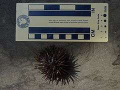
Face - aboral
-
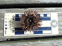
Face - oral
Habitat and range
Its natural habitat is in the western Atlantic Ocean. A. punctulata can be found in shallow water from Massachusetts to Cuba and the Yucatan Peninsula, from Texas to Florida in the Gulf of Mexico, the coast from Panama to French Guiana, and in the Lesser Antilles, usually on rocky, sandy, or shelly bottoms.[1]
Ecology and behaviour
A. punctulata is omnivorous, consuming a wide variety of prey[2] although Karlson[3] classified it as a generalized carnivore. Its galactolipids, rather than phlorotannins, act as herbivore deterrents in Fucus vesiculosus against A. punctulata.[4]
Uses in science
For more than a century, developmental biologists have valued the sea urchin as an experimental model organism. Sea urchin eggs are transparent and can be manipulated easily in the research laboratory. Their eggs can be easily fertilized and then develop rapidly and synchronously.[5][6]
For decades, the sea urchin embryo has been used to establish the chromosome theory of heredity, the description of centrosomes, parthenogenesis, and fertilization.[7][8][9] Research work during the last 30 years established such important phenomena as stable mRNA and translational control, isolation and characterization of the mitotic apparatus, and the realization that the major structural proteins of the mitotic apparatus are microtubules.[10][11] Sea urchin studies provided the first evidence of actin in nonmuscle cells.[12][13]
Arbacia punctulata is also a model organism of marine sediments toxicity[14][15] and sperm study.[16][17]
References
- ↑ Serafy, D. K., 1979: Echinoids (Echinodermata: Echinoidea). Mem. Hourglass Cruises, 5: 1 – 120.
- ↑ Sharp, D. T.; Gray, I. E. (1962). "Studies on factors affecting the local distribution of two sea urchins, Arbacia punctulata and Lytechinus variegatus". Ecology. 43: 309–313. doi:10.2307/1931986.
- ↑ Karlson, R., 1978: Predation and space utilization patterns in a marine epifaunal community. J. Exp. Mar. Biol. Ecol., 31: 225 – 239.
- ↑ Galactolipids rather than phlorotannins as herbivore deterrents in the brown seaweed Fucus vesiculosus. Michael S. Deal, Mark E. Hay, Dean Wilson and William Fenical, Oecologia, June 2003, Volume 136, Issue 1, pages 107-114, doi:10.1007/s00442-003-1242-3
- ↑ RULON O (December 1947). "The modification of developmental patterns in Arbacia eggs with malonic acid". Anat. Rec. 99 (4): 652. PMID 18895450.
- ↑ Kanungo J (June 2002). "Prolonged incubation in seawater induces a DNA-dependent protein phosphorylation activity in Arbacia punctulata eggs". Biochem. Biophys. Res. Commun. 294 (3): 667–71. doi:10.1016/S0006-291X(02)00539-9. PMID 12056821.
- ↑ FAILLA PM (June 1965). "RECOVERY FROM DIVISION DELAY IN IRRADIATED GAMETES OF ARBACIA PUNCTULATA". Radiat. Res. 25 (2): 331–40. doi:10.2307/3571975. JSTOR 3571975. PMID 14295124.
- ↑ Sachs MI, Anderson E (October 1970). "A cytological study of artificial parthenogenesis in the sea urchin Arbacia punctulata". J. Cell Biol. 47 (1): 140–58. doi:10.1083/jcb.47.1.140. PMC 2108410
 . PMID 4327513.
 - ↑ Kite GL (October 1912). "THE NATURE OF THE FERTILIZATION MEMBRANE OF THE EGG OF THE SEA URCHIN (ARBACIA PUNCTULATA)". Science. 36 (930): 562–564. doi:10.1126/science.36.930.562-a. PMID 17812420.
- ↑ ZIMMERMAN AM, MARSLAND D (July 1964). "CELL DIVISION: EFFECTS OF PRESSURE ON THE MITOTIC MECHANISMS OF MARINE EGGS (ARBACIA PUNCTULATA)". Exp. Cell Res. 35 (2): 293–302. doi:10.1016/0014-4827(64)90096-5. PMID 14195437.
- ↑ SCOTT A (October 1950). "A cytological analysis of the effects of cyanide and 4,6-dinitro-orthocresol on the mitotic phases in Arbacia punctulata". Biol. Bull. 99 (2): 362–3. PMID 14791535.
- ↑ Henson JH, Schatten G (1983). "Calcium regulation of the actin-mediated cytoskeletal transformation of sea urchin coelomocytes". Cell Motil. 3 (5-6): 525–34. doi:10.1002/cm.970030519. PMID 6420068.
- ↑ Kabat-Zinn J, Singer RH (April 1981). "Sea urchin tube feet: unique structures that allow a cytological and molecular approach to the study of actin and its gene expression". J. Cell Biol. 89 (1): 109–14. doi:10.1083/jcb.89.1.109. PMC 2111775. PMID 6894447.
- ↑ Jäntschi L, Bolboaca SD (2008). "A structural modelling study on marine sediments toxicity". Mar Drugs. 6 (2): 372–88. doi:10.3390/md20080017. PMC 2525494. PMID 18728732.
- ↑ Rudolph A, Medina P, Urrutia C, Ahumada R (July 2008). "Ecotoxicological sediment evaluations in marine aquaculture areas of Chile". Environ Monit Assess. 155 (1-4): 419–29. doi:10.1007/s10661-008-0444-x. PMID 18633720.
- ↑ Lillie FR (March 1915). "The Fertilizing Power of Sperm Dilutions of Arbacia". Proc. Natl. Acad. Sci. U.S.A. 1 (3): 156–60. doi:10.1073/pnas.1.3.156. PMC 1090763. PMID 16575966.
- ↑ Inamdar MV, Kim T, Chung YK, Was AM, Xiang X, Wang CW, Takayama S, Lastoskie CM, Thomas FI, Sastry AM (November 2007). "Assessment of sperm chemokinesis with exposure to jelly coats of sea urchin eggs and resact: a microfluidic experiment and numerical study". J. Exp. Biol. 210 (Pt 21): 3805–20. doi:10.1242/jeb.005439. PMID 17951422.