Adipose tissue macrophages
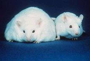
Adipose tissue macrophages (abbr. ATMs) comprise tissue resident macrophages present in adipose tissue. Adipose tissue apart from adipocytes is composed of the stromal vascular fraction (SVF) of cells including preadipocytes, fibroblasts, vascular endothelial cells and variety of immune cells. The latter ones are composed of mast cells, eosinophils, B cells, T cells and macrophages.[1] The number of macrophages within adipose tissue differs depending on the metabolic status. As discovered by Rudolph Leibel and Anthony Ferrante et al. in 2003 at Columbia University, the percentage of macrophages within adipose tissue ranges from 10% in lean mice and humans up to 50% in extremely obese, leptin deficient mice and almost 40% in obese humans.[2] Increased number of adipose tissue macrophages correlates with increased adipose tissue production of proinflammatory molecules and might therefore contribute to the pathophysiological consequences of obesity (e.g. insulin resistance, type 2 diabetes).[3]
M1/M2 macrophage polarization
Macrophages are remarkably plastic cells which in order to adapt to different tissue microenvironments can assume a range of different phenotypes. Accordingly, macrophages can exhibit either pro- or anti-inflammatory phenotypes and are routinely classified into M1 (classically activated) phenotype and M2 (alternatively activated) phenotype.[4] According to this classification, macrophages acquire M1 phenotype following in vitro stimulation with interferon gamma (IFN-γ) alone or in combination with TLR ligands (e.g. lipopolysaccharide (LPS)) whereas macrophages acquire M2 phenotype after in vitro exposure to IL-4 and IL-13. M1 macrophages secrete high levels of proinflammatory cytokines (e.g. tumor necrosis factor (TNF-α), IL-6, IL-1β) and generate reactive oxygen and nitrogen species such as nitric oxide via activation of inducible nitric oxide synthase (iNOS). Conversely, M2 macrophages activate arginase 1 (Arg1) that blocks iNOS activity and therefore inhibits nitric oxide production. They also secrete anti-inflammatory cytokines (e.g. IL-10, TGF-β, IL-4) essential for inflammatory response resolution. M1 macrophages are microbicidal and tumoricidal, and stimulate adaptive immune response. M2 macrophages are associated with anti-inflammatory and homeostatic functions linked to wound healing. However, in this classification system, M1 and M2 macrophages are regarded as two extreme phenotypes. For example, macrophages stimulated with IL-4 and IL-13 are defined as M2a, whereas macrophages stimulated with LPS and apoptotic cells as M2b and macrophages stimulated with IL-10, transforming growth factor-β (TGF-β) or glucocorticoids as M2c.[5] In adipose tissue, distinction between M1 and M2 macrophage polarization can be monitored by assessing the expression of selected markers. Macrophages displaying M1 phenotype have been characterized by expression of F4/80, CD11c and iNOS whereas macrophages displaying M2 phenotype have been characterized by expression of F4/80, CD301 and Arg1.[6]
Adipose tissue macrophages and obesity
Increased recruitment of macrophages into adipose tissue is multifactoral.[7] Adipocyte cell death observed within pathologically expanding adipose tissue is one of the factors. Macrophages are specialized phagocytes that remove dying or dead cells or cellular debris. Within adipose tissue, presence of dead adipocytes is a hallmark of obesity. Macrophages surrounding dying or dead adipocytes form crown-like structures (CLSs), identified by the absence of perilipin staining.[8]
In addition to increased number of macrophages within adipose tissue, obesity also induces a phenotypic switch in these cells toward the classically activated (M1) phenotype.[9] Moreover, expression of inflammatory cytokines such as TNF-α is mostly derived from macrophages rather than adipocytes.[10] It has been proposed that their presence contributes to the development of insulin resistance and diabetes type-2.
Adipose tissue macrophages isolated from obese patients express growth factors, cytokines, chemokines, and proteolytic enzymes involved in the regulation of tumor growth, angiogenesis, invasion, and metastatic spread, and resemble macrophages present in tumor stroma.[11]
Adipose tissue macrophages and weight loss
Acute weight loss is also associated with increased, yet transient recruitment of macrophages into adipose tissue. However the recruited macrophages do not promote inflammatory response but rather regulate lipolysis. Recruited macrophages are characterized by higher expression of scavenger receptors (i.e. CD36 and macrophage scavenger receptor 1 (MSR1)) and lipid-handling genes (i.e. adipose differentiation-related protein (Adfp), fatty acid-binding protein 4 (Fabp4), ApoE and ABCA1), and increased accumulation of Oil Red O-positive lipids. In this case, release of free fatty acids (FFAs) serves as a signal for macrophage recruitment.[12] [13]
Adipose tissue macrophages and tumor growth
Macrophages within tumor stroma, so called tumor-associated macrophages (TAMs) promote tumor growth and metastasis.[14] Tumor-associated macrophage infiltration correlates with poor prognosis in patients with breast, cervix, bladder and brain cancers.[15] Pathophysiological interaction between tumor-associated macrophages and surrounding cells, such as endothelial cells promote tumor progression. In 1971, Judah Folkman proposed that angiogenesis plays essential role in tumor growth.[16] Macrophages secrete many pro-angiogenic factors including vascular endothelial growth factor (VEGF), TNF-α, granulocyte macrophage colony-stimulating factor (GM-CSF) and IL-1 and IL-6.[17] Additionally it has been shown that adipose tissue surrounding certain tumors or metastases to the lymph nodes, which are embedded in adipose tissue, fuels tumor growth by serving as a depot for adipose tissue macrophages that stumulate angiogenesis and resemble TAMs.[18] [19] [20]
References
- ↑ Schipper HS, Prakken B, Kalkhoven E, Boes M. Adipose tissue-resident immune cells: key players in immunometabolism. Trends Endocrinol Metab 2012; 23:407-15.
- ↑ Weisberg SP, McCann D, Desai M, Rosenbaum M, Leibel RL, Ferrante AW. Obesity is associated with macrophage accumulation in adipose tissue. Journal of Clinical Investigation 2003; 112:1796-808.
- ↑ Lumeng CN, Bodzin JL, Saltiel AR. Obesity induces a phenotypic switch in adipose tissue macrophage polarization. J Clin Invest 2007; 117:175-84.
- ↑ Mosser DM, Edwards JP. Exploring the full spectrum of macrophage activation. Nat Rev Immunol 2008; 8:958-69.
- ↑ Mantovani A, Sica A, Sozzani S, Allavena P, Vecchi A, Locati M. The chemokine system in diverse forms of macrophage activation and polarization. Trends Immunol 2004; 25:677-86.
- ↑ Eagle AR, Chawla A. In obesity and weight loss, all roads lead to the mighty macrophage. Journal of Clinical Investigation 2010; 120:3437-40.
- ↑ Surmi BK, Hasty AH. Macrophage infiltration into adipose tissue: initiation, propagation and remodeling. Future Lipidol 2008; 3:545-56.
- ↑ Cinti S, Mitchell G, Barbatelli G, Murano I, Ceresi E, Faloia E, et al. Adipocyte death defines macrophage localization and function in adipose tissue of obese mice and humans. J Lipid Res 2005; 46:2347-55.
- ↑ Lumeng CN, Bodzin JL, Saltiel AR. Obesity induces a phenotypic switch in adipose tissue macrophage polarization. J Clin Invest 2007; 117:175-84.
- ↑ Weisberg SP, McCann D, Desai M, Rosenbaum M, Leibel RL, Ferrante AW. Obesity is associated with macrophage accumulation in adipose tissue. Journal of Clinical Investigation 2003; 112:1796-808.
- ↑ Mayi TH, Daoudi M, Derudas B, Gross B, Bories G, Wouters K, et al. Human adipose tissue macrophages display activation of cancer-related pathways. J Biol Chem 2012; 287:21904-13.
- ↑ Eagle AR, Chawla A. In obesity and weight loss, all roads lead to the mighty macrophage. Journal of Clinical Investigation 2010; 120:3437-40.
- ↑ Kosteli A, Sugaru E, Haemmerle G, Martin JF, Lei J, Zechner R, et al. Weight loss and lipolysis promote a dynamic immune response in murine adipose tissue. Journal of Clinical Investigation 2010; 120:3466-79.
- ↑ Biswas SK, Sica A, Lewis CE. Plasticity of macrophage function during tumor progression: regulation by distinct molecular mechanisms. J Immunol 2008; 180:2011-7.
- ↑ Bingle L, Brown NJ, Lewis CE. The role of tumour-associated macrophages in tumour progression: implications for new anticancer therapies. J Pathol 2002; 196:254-65.
- ↑ Cao YH, Langer R. A review of Judah Folkman's remarkable achievements in biomedicine. P Natl Acad Sci USA 2008; 105:13203-5.
- ↑ Lin EY, Li JF, Gnatovskiy L, Deng Y, Zhu L, Grzesik DA, et al. Macrophages regulate the angiogenic switch in a mouse model of breast cancer. Cancer Res 2006; 66:11238-46.
- ↑ Wagner M, Bjerkvig R, Wiig H, Melero-Martin JM, Lin RZ, Klagsbrun M, et al. Inflamed tumor-associated adipose tissue is a depot for macrophages that stimulate tumor growth and angiogenesis. Angiogenesis 2012; 15:481-95.
- ↑ Wagner M, Bjerkvig R, Wiig H, Dudley AC. Loss of adipocyte specification and necrosis augment tumor-associated inflammation. Adipocyte. 2013; 2:176-83.
- ↑ Wagner M, Dudley AC. A three-party alliance in solid tumors: Adipocytes, macrophages and vascular endothelial cells. Adipocyte. 2013; 2:67-73.