14-3-3 protein
| 14-3-3 | |||||||||
|---|---|---|---|---|---|---|---|---|---|
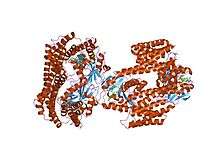 Crystal structure of the 14-3-3 zeta:serotonin N-acetyltransferase complex.[1] | |||||||||
| Identifiers | |||||||||
| Symbol | 14-3-3 | ||||||||
| Pfam | PF00244 | ||||||||
| InterPro | IPR000308 | ||||||||
| SMART | 14_3_3 | ||||||||
| PROSITE | PDOC00633 | ||||||||
| SCOP | 1a4o | ||||||||
| SUPERFAMILY | 1a4o | ||||||||
| |||||||||
14-3-3 proteins are a family of conserved regulatory molecules that are expressed in all eukaryotic cells. 14-3-3 proteins have the ability to bind a multitude of functionally diverse signaling proteins, including kinases, phosphatases, and transmembrane receptors. More than 200 signaling proteins have been reported as 14-3-3 ligands.
The name 14-3-3 refers to the particular elution and migration pattern of these proteins on DEAE-cellulose chromatography and starch-gel electrophoresis. The 14-3-3 proteins eluted in the 14th fraction of bovine brain homogenate and were found on positions 3.3 of subsequent electrophoresis by Moore and Perez (1967).
Elevated amounts of 14-3-3 proteins are found in the cerebrospinal fluid of patients with Creutzfeldt–Jakob disease.[2]

Properties of 14-3-3 proteins
There are seven genes that encode seven distinct 14-3-3 proteins in most mammals (See Human genes below) and 13-15 genes in many higher plants, though typically in fungi they are present only in pairs. Protists have at least one. Eukaryotes can tolerate the loss of a single 14-3-3 gene if multiple genes are expressed, however deletion of all 14-3-3s (as experimentally determined in yeast) results in death.
14-3-3 proteins can be considered evolved members of the Tetratrico Peptide Repeat (TPR) superfamily, generally have 9 or 10 alpha helices, and usually form homo- and/or hetero-dimer interactions along their amino-termini helices. These proteins contain a number of known common modification domains, including regions for divalent cation interaction, phosphorylation & acetylation, and proteolytic cleavage, among others established and predicted.
There are common recognition motifs for 14-3-3 proteins that contain a phosphorylated serine or threonine residue; Mode 1 is R[SFYW]XpSXP & Mode 2 RX[SYFWTQAD]Xp(S/T)X[PLM] (where an 'x' can be several, but not any of the 20 amino acids; a lower case 'p' indicates the site of phosphorylation) but also binding to non-phosphorylated ligands has been reported. This interaction occurs along a so-called binding groove or cleft that is amphipathic in nature. To date, the crystal structures of six classes of these proteins have been resolved and deposited in the public domain.
Function
14-3-3 proteins play an isoform-specific role in class switch recombination. They are believed to interact with the protein Activation-Induced (Cytidine) Deaminase in mediating class switch recombination.
Phosphorylation of Cdc25C by CDS1 and CHK1 creates a binding site for the 14-3-3 family of phosphoserine binding proteins. Binding of 14-3-3 has little effect on Cdc25C activity, and it is believed that 14-3-3 regulates Cdc25C by sequestering it to the cytoplasm, thereby preventing the interactions with CycB-Cdk1 that are localized to the nucleus at the G2/M transition.[3]
The eta isoform is reported to be a biomarker (in synovial fluid) for rheumatoid arthritis.[4]
14-3-3 regulating cell-signalling
Human genes
- YWHAB - "14-3-3 beta"
- YWHAE - "14-3-3 epsilon"
- YWHAG - "14-3-3 gamma"
- YWHAH - "14-3-3 eta"
- YWHAQ - "14-3-3 tau"
- YWHAZ - "14-3-3 zeta"
- SFN or YWHAS - "14-3-3 sigma"
The 14-3-3 proteins alpha and delta (YWHAA and YWHAD) were found to be equivalent to YWHAB and YWHAZ, respectively.
14-3-3 in plants
Presence of large gene families of 14-3-3 proteins in the Viridiplantae kingdom reflects their essential role in plant physiology. A phylogenetic analysis of 27 plant species clustered the 14-3-3 proteins into four groups.
14-3-3 proteins activate the auto-inhibited plasma membrane P-type H+ ATPases. They bind the ATPases' C-terminus at a conserved threonine.[6]
Further reading
- Moore BW, Perez VJ (1967). FD Carlson, ed. Physiological and Biochemical Aspects of Nervous Integration. Prentice-Hall, Inc., The Marine Biological Laboratory, Woods Hole, MA. pp. 343–359.
- Mhawech P (Apr 2005). "14-3-3 proteins--an update". Cell Research. 15 (4): 228–236. doi:10.1038/sj.cr.7290291. PMID 15857577.
- Steinacker P, Aitken A, Otto M (Sep 2011). "14-3-3 proteins in neurodegeneration". Seminars in Cell & Developmental Biology. 22 (7): 696–704. doi:10.1016/j.semcdb.2011.08.005. PMID 21920445.
References
- ↑ T. Obsil; R. Ghirlando; D. C. Klein; S. Ganguly & F. Dyda (April 2001). "Crystal structure of the 14-3-3zeta:serotonin N-acetyltransferase complex. a role for scaffolding in enzyme regulation". Cell. 105 (2): 257–267. doi:10.1016/S0092-8674(01)00316-6. PMID 11336675.
- ↑ Takahashi H, Iwata T, Kitagawa Y, Takahashi RH, Sato Y, Wakabayashi H, Takashima M, Kido H, Nagashima K, Kenney K, Gibbs CJ, Kurata T (Nov 1999). "Increased levels of epsilon and gamma isoforms of 14-3-3 proteins in cerebrospinal fluid in patients with Creutzfeldt–Jakob disease". Clinical and Diagnostic Laboratory Immunology. 6 (6): 983–5. PMC 95810
 . PMID 10548598.
 - ↑ Cann KL, Hicks GG (Dec 2007). "Regulation of the cellular DNA double-strand break response". Biochemistry and Cell Biology = Biochimie Et Biologie Cellulaire. 85 (6): 663–74. doi:10.1139/O07-135. PMID 18059525.
- ↑ Detection of high levels of 2 specific isoforms of 14-3-3 proteins in synovial fluid from patients with joint inflammation.
- ↑ Saha, Madhurima; Carriere, Audrey; Cheerathodi, Mujeeburahiman; Zhang, Xiaocui; Lavoie, Geneviève; Rush, John; Roux, Philippe; Ballif, Bryan (2012). "RSK phosphorylates SOS1 creating 14-3-3-docking sites and negatively regulating MAPK activation". Biochemical Journal. 447: 159–66. doi:10.1042/BJ20120938. PMC 4198020. PMID 22827337.
- ↑ Jahn TP, Schulz A, Taipalensuu J, Palmgren MG (Feb 2002). "Post-translational modification of plant plasma membrane H(+)-ATPase as a requirement for functional complementation of a yeast transport mutant". The Journal of Biological Chemistry. 277 (8): 6353–6358. doi:10.1074/jbc.M109637200. PMID 11744700.
External links
- Eukaryotic Linear Motif resource motif class LIG_14-3-3_1
- Eukaryotic Linear Motif resource motif class LIG_14-3-3_2
- Eukaryotic Linear Motif resource motif class LIG_14-3-3_3
- 14-3-3 Protein at the US National Library of Medicine Medical Subject Headings (MeSH)
- Three-dimensional structure of 14-3-3 Protein Theta (Human) complexed with a peptide in the PDB.
- Drosophila 14-3-3epsilon - The Interactive Fly
- Drosophila 14-3-3zeta - The Interactive Fly